3-D CT Scans (Cone Beam) for Precise Oral Surgery
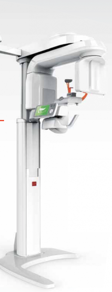
 Accurate and detailed imaging is integral to performing any surgery, including oral and maxillofacial surgery. Advancements in imaging have helped improve the safety and efficiency of oral surgery for patients. The 3-D CT scan, or cone-beam imaging, is one of these advanced options for higher precision in planning and performing oral surgery. This technology is available at Torrance Oral Surgery and Dental Implant Center.
What is a 3-D Cone-Beam CT Scan?
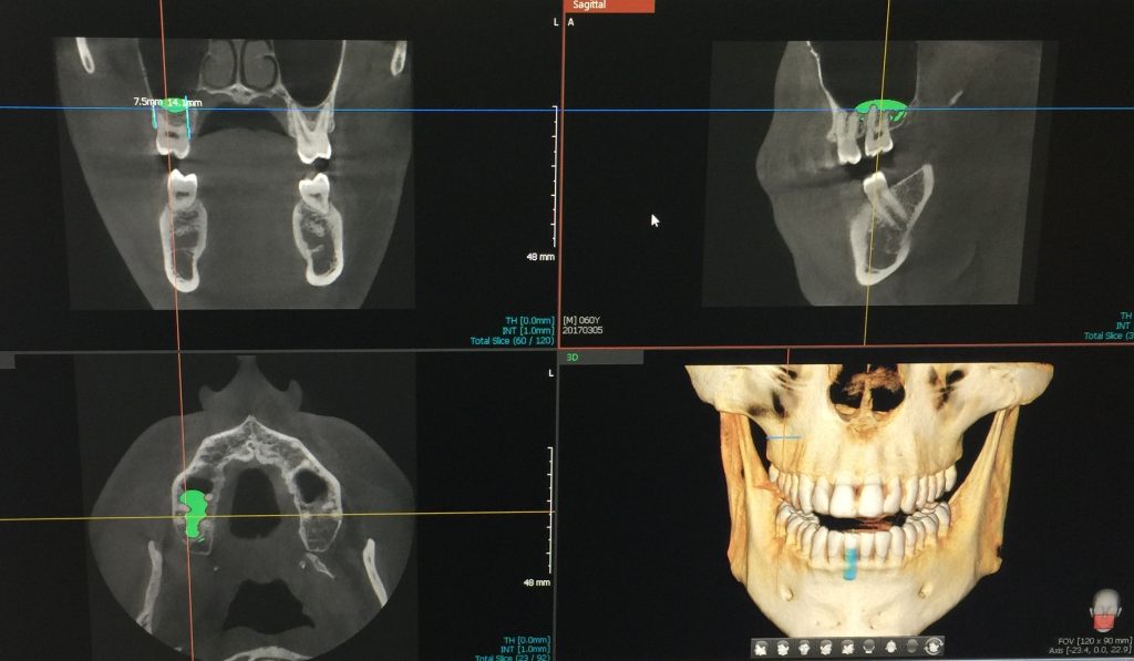
When an x-ray is performed, it takes a two-dimensional, or 2D, image of the area, showing the outline of bone, teeth and other internal structures. For many procedures, this is all that is needed. However, when intricate oral surgery will be performed, seeing the exact positions of bone, teeth and tissues in a three-dimensional image allows for a more precise view. This allows the oral surgeon to provide a more accurate diagnosis for oral conditions, as well as plan and perform complex surgeries.
The use of 3-D cone-beam CT (computed tomography) creates a three-dimensional image of the treatment area with advanced precision in detail. This is a digital image that shows the exact formations to view abnormalities and help oral surgeons plan their treatment or surgical procedure. In addition to offering a more advanced view of the treatment area, cone-beam technology is lower in radiation exposure than many other types of imaging. Both the upper and lower jaw can be scanned at one time, making it quick and convenient for the patient and medical team.
It is a top priority for Dr. Benjamin Yagoubian to have the most advanced equipment and technology at our clinic. This improves the level of accuracy and safety of our procedures, so we can give our patients the best care. We use 3-D cone-beam CT scans, updated medical technology and use the latest techniques in oral surgery help us provide excellence in oral and maxillofacial surgery for our patients.


